Sample deliverable — CITE-seq (protein + RNA)
Antibody-based protein measurement alongside RNA has its own quality-control requirements, and applying the plain single-cell recipe to it misses the ones that matter. This is a full run on public data (10x Genomics 5k PBMC with TotalSeq-B (public CITE-seq)).
Do this on your own data: protein and RNA disagree in real data, and which of the two you believe has to be decided on your own panel. Upload the RNA half and the free QC report returns the per-sample cut-offs and the expected doublet count: omics.hstgenomics.com/qc-check. The checks a CITE-seq delivery is supposed to contain: omics.hstgenomics.com/guides/citeseq-what-to-check.
Want it done rather than shown? We do analysis only — send a count matrix, an h5ad or a Seurat object, Cell Ranger output or a public accession and get the analysis back with the code that produced it, at a fixed price agreed before any work starts: omics.hstgenomics.com/quote.
1 · Isotype controls — the check that decides whether anything is real
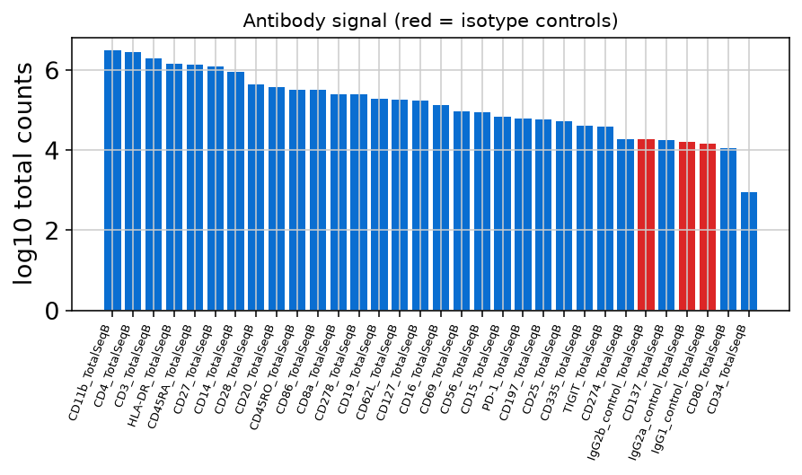
Isotype controls are antibodies that should not bind anything; whatever they register is non-specific background. In this dataset they carry 0.25% of the total antibody signal, which is low — so a positive stain here means something. Without this number, every "positive" population in a CITE-seq figure is unverified. It is the first thing we compute and the first thing we report.
2 · Antibody counts are normalised differently from RNA
CLR (centred log-ratio per cell), as recommended for antibody-derived tags. Antibody-derived counts have a different noise structure from transcripts — a few antibodies dominate the library and staining efficiency varies between markers — so the RNA normalisation is not appropriate for them.
3 · Protein against RNA, cluster by cluster
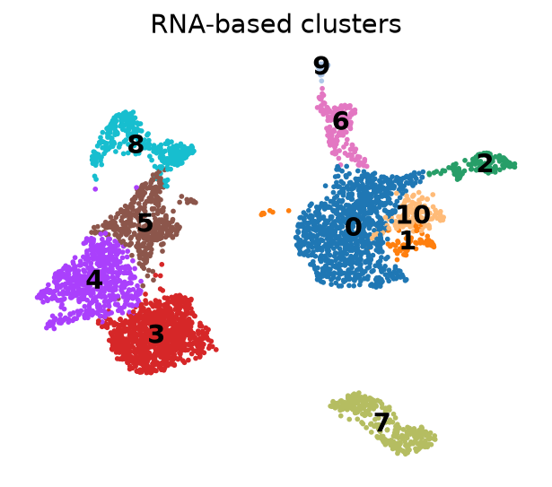
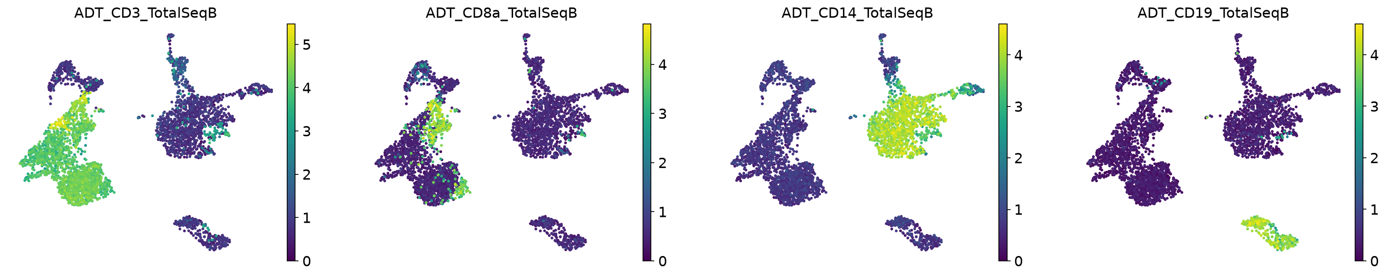
Two independent measurements of the same cells should point at the same populations. For each canonical pair we compare the antibody signal with its RNA counterpart across RNA-defined clusters — 5 of 5 agree on which cluster is highest.
| Antibody | RNA gene | Correlation across clusters | Peak cluster |
|---|---|---|---|
CD3_TotalSeqB | CD3E |
0.935 | ✔ same cluster |
CD19_TotalSeqB | MS4A1 |
0.999 | ✔ same cluster |
CD14_TotalSeqB | CD14 |
0.968 | ✔ same cluster |
CD8a_TotalSeqB | CD8A |
0.998 | ✔ same cluster |
CD4_TotalSeqB | IL7R |
0.543 | ✔ same cluster |
4 · Where they disagree is biology, not error
CD3, CD19, CD14 and CD8a track their transcripts almost perfectly
(r ≥ 0.93). CD4 does not — correlation with IL7R is only
0.543. That is expected: CD4 protein
is present on monocytes as well as helper T cells, while IL7R is a T-cell transcript, and protein
persists long after its mRNA is gone. A report that treats this mismatch as an error has misread the assay; a
report that does not notice it has not checked.
What a full CITE-seq project adds
- Background subtraction using the isotype controls, with the residual reported per antibody.
- Joint clustering on RNA and protein (WNN or equivalent), compared against RNA-only clustering.
- Gating-style population definitions that a flow-cytometry-trained reader can follow.
- Differential abundance and expression at subject level, as in any other design.
- The script, so it can be re-run: see the code.